The Cavernous Sinus
Pass USMLE Step 1, Step 2 CK and Step 3. Only the Best USMLE Prep Program provides the Best USMLE Review Course Content in this rapidly expanding universe of medical information. Become conversant with the new medical discoveries that is transforming our world.
At USMLE Insider Prep Course, we use powerful, illustrative imagery to teach you the challenging concepts that must be mastered for USMLE success. The corticobulbar tract is a poorly understood concept for most medical students. But we make this stuff ridiculously easy. This is what separates USMLE Insider from all the other USMLE Step 1 review courses.
The cavernous sinus, one of the dural venous sinuses, is a particularly important structure. It has relationships with many structures with great clinical significance for the USMLE. Before discussing the clinical importance of the cavernous sinus, a review of the dural venous sinuses is necessary:
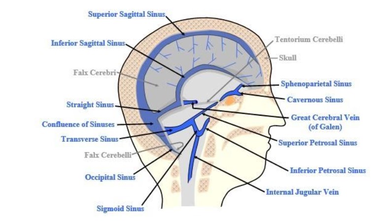
AN OVERVIEW OF THE DURAL VENOUS SINUSES
-
Formed primarily within folds of dura mater.
-
They are superior sagittal, inferior sagittal, straight, transverse, sigmoid, cavernous, superior and inferior petrosal sinuses, occipital and sphenoparietal sinus.
-
The superior sagittal, straight and inferior sagittal sinuses drain into the confluence of sinuses.
-
The inferior petrosal sinus drains the cavernous sinus and then drains into the internal jugular vein.
-
The superior ophthalmic veins and sphenoparietal sinus drain into the cavernous sinus.
-
The superior petrosal sinus also drains the cavernous sinus and empty into the sigmoid sinus at its junction with the transverse sinus.
-
The transverse sinuses subserve the function of draining the confluence of the sinuses. They also form the sigmoid sinuses.

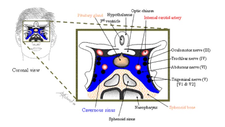
Figure 1. Two views of the cavernous sinus: sagittal and coronal.
Now, some important clinical correlations of the cavernous sinus:
CRANIAL NERVES:
The cranial nerves in the cavernous sinus are:
-
CN VI (which lies inside the sinus near the internal carotid artery) and
-
CN III, IV, V1, and V2 (all of which lie in the lateral walls of the cavernous sinus).
-
It is especially important to remember that the mandibular nerve is the only branch of the trigeminal that has no relationship with the cavernous sinus.
The clinical significance is as follows:
-
The abducens nerve lies inside the cavernous sinus alongside the internal carotid artery. For this reason, this nerve is the first to be compressed by internal carotid artery thrombosis, fistula, or aneurysm.
-
Cavernous sinus thrombosis would cause deficits related to structures in the cavernous sinus, which are CN III, IV, V1, V2, CN VI, and the Horner's oculosympathetic fibers.
-
Imagine a tumor of the pituitary hypophysis that encroaches the cavernous sinus; if such a tumor extends laterally, the first nerve to suffer is CN VI because it is the most medial.
PITUITARY STALK:
Notice the inferior position of the pituitary gland relative to the optic chiasm, which is not within the sinus. If a pituitary tumor extends anterosuperiorly, the nerve that will be compressed will be optic chiasm, resulting in bitemporal hemianopsia.
TRANSPHENOIDAL APPROACH:
To treat pituitary adenomas, administration of dopamine agonists such as bromocriptine is the gold standard, because dopamine is the prolactin-inhibiting factor. Surgery is also an option: the hypophysis is approached transnasally through the sphenoid sinus - the so-called transphenoidal approach of neurosurgery.
Observe the relationship of the nose, the sphenoid bone, sphenoidal sinus and the pituitary gland in the illustration below.
EMPTY SELLA SYNDROME:
In this illustration below, we place special emphasis on the diaphragma sellae, a membrane that sits on top of the pituitary and protects it in its socket within the bones at the base of the skull.
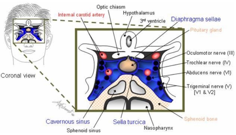
With a large opening in the diaphragma sellae, CSF will flow into the pituitary area and flatten the pituitary out within the sellae. This is called empty sella syndrome. Generally, the pituitary is perfectly normal; it is just not seen on radiographs because of the spinal fluid covering it. There is usually a normal level of pituitary hormones, but some patients may present with headache and visual problems.
THE 'DANGER AREA' OF THE FACE:
The origin of the facial vein, the angular vein, communicates with the cavernous sinus by way of the ophthalmic veins. In the cheek, the facial vein receives the deep facial vein from the pterygoid plexus, and it usually ends directly or indirectly in the internal jugular vein. Because of its connections with the cavernous sinus and the pterygoid plexus and the consequent possibility of spread of infection, the territory of the facial vein around the nose and upper lip has been termed the 'danger area' of the face.

Infections involving the periorbital space (i.e., eyelid and portions of skin around the eye, anterior to the orbital septum) can penetrate through the ophthalmic vein into the cavernous sinus and into the dural space, leading to meningitis. It may be caused by breaks in the skin around the eye; sinusitis; or from spread of infection elsewhere. Thus, prompt antibiotic therapy is critical with this infection.
Be prepared to answer questions on the probable route of spread of such infection to the brain. It goes through the facial vein to ophthalmic vein to cavernous sinus into the dura space. Thrombosis of the cavernous sinus is a dangerous condition.
CAVERNOUS SINUS THROMBOSIS:
Cavernous sinus thrombosis occurs as a complication of a contiguous spread of infection from a nasal furuncle, which accounts for 50% of cases. It may also result from spreading infection from sphenoidal or ethmoidal sinuses (30%) and dental infections (10%).
The characteristic findings include chemosis (edema of the conjunctiva), exophthalmos, headaches, and paralysis of cranial nerves and other structures which course the cavernous sinus. The cranial nerve palsies include II, III, IV, V1, V2, and VI (sixth nerve palsy is the most common). Because the oculosympathetic pathways also course through the sinus, the characteristic Horner syndrome symptoms of ptosis, miosis, and anhidrosis may be present.
The paranasal sinuses are rich with anastomotic venous system which allows retrograde spread of infection to the cavernous sinus via the superior and inferior ophthalmic veins. The ophthalmic and facial veins were previously thought to be valveless, and the absence of valves was thought to be the major cause of the retrograde spread. However, a recent study has found that the aforementioned veins are not valveless. Staphylococcus aureus is the most common bacteria involved.
Now, check out one of the USMLE questions we have prepared on these concepts. At USMLE Insider Prep Course, we have our experts stimulate your mind with challenging questions. Not only are our USMLE course notes one to die for, but we create the fertile atmosphere for your brilliant mind to flourish. We stimulate you to think critically and help you not only achieve the highest score on the USMLE but also to be a great clinician. Test yourself with this and let us see how you do. We will discuss the answer and offer explanations later.
1. A 36-year-old baseball player is struck in the left temple by an errant throw and momentarily loses consciousness. When he regains consciousness, he is taken to the emergency room for evaluation. CT scan of the head reveals an incidental aneurysm of the left internal carotid artery in the cavernous sinus projecting into the pituitary fossa. No further intracranial anomaly is identified. Which of the following cranial nerves is typically the first to be affected by such an aneurysm?
A. CN II
B. CN III
C. CN IV
D. CN VI
E. CN XI